Posterior sclera, choroidea, retina
 |
|
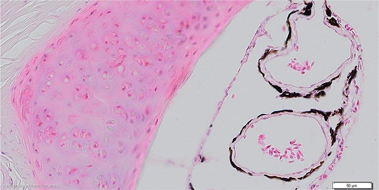
Transverse section through the sclera and choroidea of the eye of a pigeon (HE)
The choroid can be divided in: the suprachoroidea (lamina suprachoroidea), a stromal layer (lamina vasculosa) with prominent blood vessels, the choriocapillaris (lamina choroidocapillaris) and Bruch’s membrane (lamina basalis choroidea). The suprachoroidea consists of large lacunae and the membrana fusca, a pigmented tissue layer between the sclera and lacunae.
At the micrograph above, the lamina suprachoroidea is destroyed (visible as one big artefact), the membrana fusca cannot be seen and Bruch’s membrane is not present (attached to the retinal layer).