Herbst corpuscles
 |
|
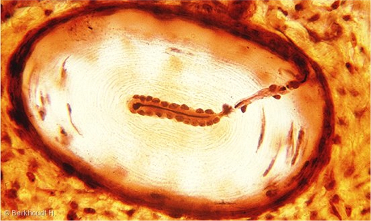
Longitudinal section of a Herbst corpuscle in the beak of a mallard (frozen section, silver staining: modification of the Bielschowsky-Gross silver impregnation technique, Berkhoudt 1980) photo courtesy of dr. H. Berkhoudt
A single myelinated afferent nerve fiber enters each Herbst corpuscle and loses its myelin sheath before it enters the inner core. At the picture above, the progress of the axon is clearly visible.
In the inner core, schwann cells are organized in twin rows around the central axon and form interdigitating lamellae. Fingerlike protrusions of the axon form contact with these lamellae, but these protrusions are not visible an light microscopic images. Often, two excentrally lying additional nuclei are present at the enlarged tip of the axon.
